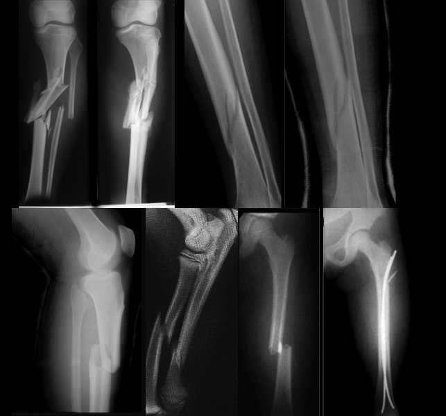
A fracture, or discontinuity of the bone, is the most common type of bone lesion. Normal bone can withstand considerable compression and shearing forces and, to a lesser extent, tension forces. A fracture occurs when more stress is placed on the bone than it is able to absorb.
A bone fracture is a medical condition in which there is a break in the continuity of the bone. A bone fracture can be the result of high force impactor stress, or trivial injury as a result of certain medical conditions that weaken the bones, such asosteoporosis, bone cancer, or osteogenesis imperfecta, where the fracture is then termedpathological fracture.
(http://en.wikipedia.org/wiki/Bone_fracture)
Cause for Bone Fractures
Bone Fractures Grouped according to cause, fractures can be divided into three major categories:
- Fractures caused by sudden injury The most common fractures result from major trauma, such as a fall on an outstretched arm, a skiing or motor vehicle accident, and child, spouse, or elder abuse (shown by multiple or repeated episodes of fractures). The force causing the fracture may be direct, such as a fall, or indirect, such as a massive muscle contraction or trauma transmitted along the bone. For example, the head of the radius or clavicle can be fractured by the indirect forces that result from falling on an outstretched hand.
- Fatigue or stress fractures A fatigue fracture results from repeated wear on a bone. Pain associated with overuse injuries of the lower extremities, especially posterior medial tibial pain, is one of the most common symptoms that physically active persons, such as runners, experience Stress fractures in the tibia.
- Pathologic fractures. A pathologic fracture is fracture that occurs when the normal integrity and strength of bone have been compromised by invasive disease or destructive processes or tumors. Fractures of this type may occur spontaneously with little or no stress. The underlying disease state can be local, as with infections, cysts, or tumors, or it can be generalized, as in osteoporosis, Paget’s disease, or disseminated tumors.
Classification of Bone Fractures
Fractures usually are classified according to:
Location
Type
Direction or pattern of the fracture line.
Fragment position
- Angulated, Bone fragments are at an angle to each other
- Avulsed, Bone fragments are pulled from normal position by muscle spasms, muscle contractions, or ligament resistance
- Comminuted, Bone breaks into many small pieces
- Displaced, Bone fragments separate and are deformed Impacted, A bone fragment is forced into another bone or bone fragment
- Nondisplaced, After the fracture, two sections of the bone maintain normal alignment
- Overriding, Bone fragments overlap, thereby shortening the total length of the bone
- Segmental
Fracture line
- Linear Fracture line is parallel to the axis of the bone
- Longitudinal Fracture line extends longitudinally but not parallel to the axis of the bone
- Oblique Fracture line crosses the bone at a 45-degree angle to the axis of the bone
- Spiral Fracture line coils around the bone
- Transverse Fracture line forms a 90-degree angle to the axis of the bone
A fracture of the long bone is described in relation to its position in the boneproximal, midshaft, and distal. Other descriptions are used when the fracture affects the head or neck of a bone, involves a joint, or is near a prominence such as a condyle or malleolus. The type of fracture is determined by its communication with the external environment, the degree of break in continuity of the bone, and the character of the fracture pieces.10
A fracture can be classified as open or closed. When the bone fragments have broken through the skin, the fracture is called an open or compound fracture. In a closed fracture, there is no communication with the outside skin.
The degree of a fracture is described in terms of a partial or complete break in the continuity of bone. A greenstick fracture, which is seen in children, is an example of a partial break in bone continuity and resembles that seen when a young sapling is broken. This kind of break occurs because children’s bones, especially until approximately 10 years of age, are more resilient than the bones of adults.
The character of the fracture pieces may also be used to describe a fracture. A comminuted fracture has more than two pieces. A compression fracture, as occurs in the vertebral body, involves two bones that are crushed or squeezed together. A fracture is called impacted when the fracture fragments are wedged together. This type usually occurs in the humerus, often is less serious, and usually is treated without surgery. Segmental fracture Bone fractures occur in two areas next to each other with an isolated section in the center
The direction of the trauma produces a certain configuration or pattern of fracture. Reduction is the restoration of a fractured bone to its normal anatomic position. The pattern of a fracture indicates the nature of the trauma and provides information about the easiest method for reduction. Linear fractures, Fracture line is parallel to the axis of the bone Transverse fractures are caused by simple angulatory forces. A spiral fracture results from a twisting motion, or torque. A transverse fracture is not likely to become displaced
Treatment for Bone Fractures
The primary goals of treatment are to return the injured limb to maximal function, to prevent complications, and to obtain the best possible cosmetic results. Emergency treatment consists of splinting the limb above and below the suspected fracture where it lies, applying a cold pack, and elevating the limb, all of which reduce edema and pain. A severe fracture that causes blood loss calls for direct pressure to control bleeding. The patient with a severe fracture may also need fluid replacement (including blood products) to prevent or treat hypovolemic shock.
Treatment Options for Bone Fractures
Treatment Options to set a Bone Fractures depends on the location and severity of the injury. To heal a bone fractures properly, the fractured bone must be realigned. The most common realignment procedures are:
Immobilization using a cast or splint
Setting of bone through surgery. Advantages of surgery include: early mobility of injured bone and some use of the injured bone within weeks rather than months.
After the bone is realigned properly, medication and rehabilitation will help the recovery process. Medication is used to lessen the pain. Rehabilitation prevents stiffness. Rehabilitation involves light movement of the tissues surrounding the injury. It helps increase blood flow which will aid the healing process.
Nonoperative Management
Until comparatively recently, nonoperative treatment was the only method of treating fractures and severe soft tissue injuries, but the introduction of anesthesia, antibiotics, improved surgical implants, and better operative techniques has changed the treatment of many fractures. The process of change continues, and probably fewer fractures will be managed nonoperatively as the functional benefits of operative treatment become more apparent to both surgeons and patients.
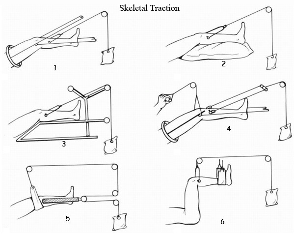
TRACTION: Skeletal Traction, Spinal Traction,
CASTS Braces
Slings, Bandages, and Support Strapping
skeletal traction
skeletal traction
Operative Management
When closed reduction is impossible, open reduction during surgery use to reduces and immobilizes the fracture by means of rods, plates, or screws
- Plating
- Intramedullary Nailing
- Kirschner wires
- External Fixation
- Arthroplasty
- Amputations
Complications for Bone Fractures
Possible complications of fractures include arterial damage, nonunion, fat embolism, infection, shock, avascular necrosis, and peripheral nerve damage. Acute Compartment Syndrome Nonunions and Bone Defects
Nursing diagnosis for bone fractures
Nursing diagnosis for bone fractures determine by data that we found in nursing assessment:
Nursing Assessment nursing care plans for bone fractures
Assessment on patient’s history usually reveals what caused the fracture, major trauma, such as a fall on an outstretched arm, a skiing or motor vehicle accident, or elder abuse. The patient typically reports pain that increases with movement and an inability to move the part of the arm or leg distal to the injury. The severity of the pain depends on the fracture type. The patient may also complain of a tingling sensation distal to the injury, possibly indicating nerve and vessel damage.
Inspection may disclose soft-tissue edema, an obvious deformity or shortening of the injured limb, and discoloration over the fracture site. Open fractures produce an obvious skin wound and bleeding. Gentle palpation usually reveals warmth, crepitus and, possibly, dislocation. Numbness distal to the injury and cool skin at the end of the extremity may indicate nerve and vessel damage.
Auscultation may reveal loss of pulses distal to the injury, an indication of possible arterial compromise or nerve damage.
Palpation pulses in distal of the fracture to detect injury to blood vessels, which is a surgical emergency
Diagnostic tests for Bone Fractures
Anteroposterior and lateral X-rays of the suspected fracture, as well as X-rays of the joints above and below it, confirm the diagnosis.
Angiography can reveal concurrent vascular injury.
MRI or CT Scan of spine if suspect a bone tumor or compression of spinal cord
Bone densitometry can predict an increased risk of osteoporosis usually in pathologic fractures
Blood tests
Nursing Care Plans for Bone Fractures. Common nursing diagnosis for bone fractures:
- Acute pain,
- Anxiety,
- Bathing or hygiene self-care deficit,
- Fear,
- Impaired physical mobility,
- Ineffective coping,
- Ineffective role performance,
- Ineffective tissue perfusion: Peripheral,
- Risk for deficient fluid volume,
- Risk for disuse syndrome,
- Risk for infection,
- Risk for injury,
- Risk for [additional] Trauma.
Nursing Goals Nursing Care Plans for Bone Fractures
Pain controlled.
Prevented or minimized Complications
Fracture stabilized.
Condition, prognosis, and therapeutic regimen understood.
Plan in place to meet needs after discharge.
Nursing Care Plans for Bone Fractures with nursing diagnosis Acute pain
Sample Nursing Care Plans For bone Fracture with nursing diagnosis Pain Acute
NURSING
DIAGNOSIS
|
INTERVENTIONS
|
EVALUATION
|
Acute Pain related to Muscle
spasms Movement of bone fragments, edema, and injury to the soft tissue
Traction, immobility device
Stress, anxiety
|
· Perform a comprehensive assessment of pain including
location, characteristics, onset/duration, frequency, quality, severity
·
Maintain
immobilization of affected part
·
Elevate
and support injured extremity
·
Perform
and supervise passive or active ROM exercises
·
Suggest diversional
activities appropriate for client’s age, physical abilities, and personal
preferences
·
Administer
medications, as indicated.
|
· Verbalize
relief of pain.
· Follow
prescribed pharmacologic regimen
· Display
relaxed manner, able to participate in activities, and sleep and rest
appropriately
· Demonstrate use of relaxation skills and diversional
activities, as indicated for individual situation
|
Patient Teaching Discharge and Home Healthcare Guidelines for Fractures
Patient
Teaching Discharge and Home Health care Guidelines for fractures patient. To prevent complications of prolonged immobility, encourage the patient to participate in physical and occupational therapy as prescribed. Verify that the patient has demonstrated safe use of assistive devices such as wheelchairs, crutches, walkers, and transfers. Teach the patient the purpose, dosage, schedule, precautions, and potential side effects, interactions, and adverse reactions of all prescribed medications. Review with the patient all follow-up appointments that are arranged. If home care is necessary, verify that appropriate arrangements have been completed.
- Help the patient set realistic goals for recovery.
- Show the patient how to use his crutches properly.
- Tell the patient with a cast to report immediately signs of impaired circulation (skin coldness, numbness, tingling, or discoloration).
- Warn the patient against getting the cast wet, and instruct him not to insert foreign objects under the cast. Teach the patient to exercise joints above and below the cast as ordered.
- Tell the patient not to walk on a leg cast or foot cast without the physician’s permission.
- Emphasize the importance of returning for follow-up care.


0 comments:
Post a Comment